Staging of Cancer: TNM System and Other Staging Methods
1. Introduction
Cancer staging is a critical process in oncology that determines the extent of cancer spread in the body. Accurate staging helps in:
- Selecting appropriate treatment plans.
- Predicting prognosis.
- Comparing treatment outcomes across populations.
There are multiple staging systems, with the TNM system being the most widely used globally. Other staging methods include the number staging system, SEER staging, and FIGO staging (for gynecological cancers).
2. The TNM Staging System
The TNM system, developed by the American Joint Committee on Cancer (AJCC) and the Union for International Cancer Control (UICC), categorizes cancer based on:
- T (Tumor size and extent of invasion)
- N (Lymph node involvement)
- M (Metastasis to distant organs)
This system is primarily used for solid tumors (e.g., breast, lung, colorectal cancers).
2.1 T-Category (Tumor Size and Local Spread)
- TX: Primary tumor cannot be assessed.
- T0: No evidence of a primary tumor.
- Tis: Tumor in situ (pre-invasive cancer, confined to original tissue).
- T1-T4: Increasing size and/or invasion into deeper structures.
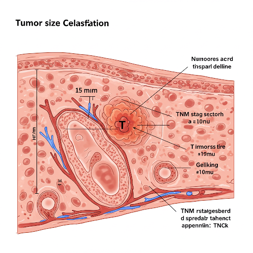
Image: Tumor Size and T Classification
(A diagram illustrating tumor invasion in different T stages.)
2.2 N-Category (Lymph Node Involvement)
- NX: Regional lymph nodes cannot be assessed.
- N0: No regional lymph node involvement.
- N1-N3: Increasing involvement of nearby lymph nodes.
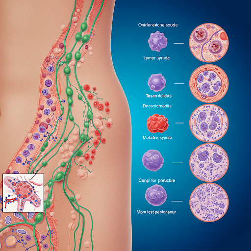
Image: Lymph Node Involvement in Cancer
(A diagram showing lymph node spread in different cancer stages.)
2.3 M-Category (Distant Metastasis)
- M0: No distant metastasis.
- M1: Distant metastasis present.
2.4 Example: TNM Staging in Breast Cancer
- T2 N1 M0 → Tumor is 2-5 cm, involves nearby lymph nodes, but no distant spread.
- T4 N3 M1 → Large tumor, extensive lymph node involvement, and metastasis present.
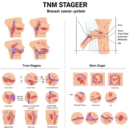
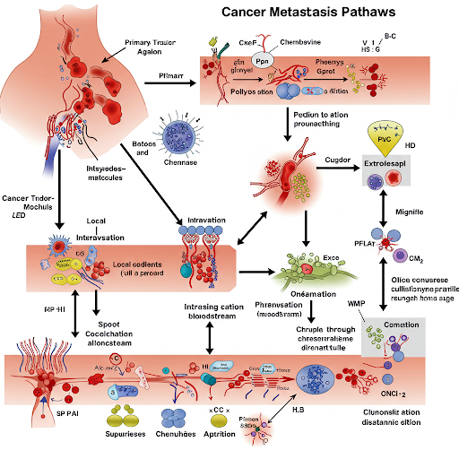
Image: Cancer Metastasis Pathways
(A visual representation of common metastatic pathways in cancer.)
Image: TNM Staging of Breast Cancer
(A stepwise representation of breast cancer progression from T1N0M0 to T4N3M1.)
3. Other Staging Methods
3.1 Number Staging System (Stages 0-IV)
This simpler system categorizes cancers into five stages:
- Stage 0: Carcinoma in situ (pre-cancerous, non-invasive).
- Stage I: Small tumor, localized, no lymph node involvement.
- Stage II: Larger tumor or spread to nearby nodes.
- Stage III: More extensive lymph node involvement.
- Stage IV: Distant metastasis (advanced cancer).
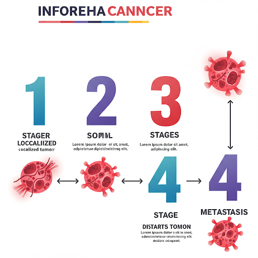
Image: Number Staging of Cancer
(A graphical depiction of cancer progression from Stage 0 to Stage IV.)
3.2 SEER Staging (Surveillance, Epidemiology, and End Results)
Used by the National Cancer Institute (NCI) for population-based cancer statistics. Categorizes cancer as:
- Localized: Confined to the organ of origin.
- Regional: Spread to nearby lymph nodes or tissues.
- Distant: Metastasized to distant organs.
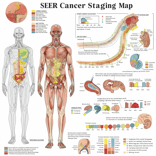
Image: SEER Cancer Staging Map
(A color-coded diagram showing cancer spread in SEER staging.)
3.3 FIGO Staging (Gynecologic Cancers)
Used for cervical, ovarian, uterine cancers, developed by the International Federation of Gynecology and Obstetrics (FIGO).
- Stage I: Limited to the organ (e.g., cervix, uterus).
- Stage II: Spread beyond the organ but not to distant sites.
- Stage III: Local lymph node involvement or peritoneal spread.
- Stage IV: Distant metastasis.
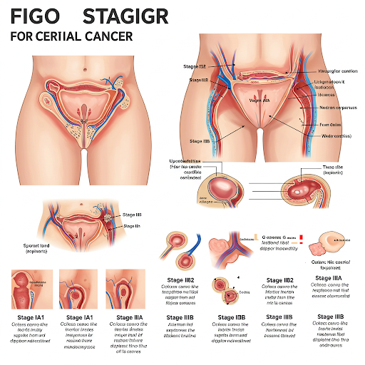
Image: FIGO Staging of Cervical Cancer
(A stepwise visual representation of cervical cancer progression.)
3.4 Ann Arbor Staging (Lymphomas)
Used for Hodgkin’s and Non-Hodgkin’s Lymphoma, based on lymph node involvement and spread.
- Stage I: Single lymph node region.
- Stage II: Multiple nodes on the same side of the diaphragm.
- Stage III: Nodes on both sides of the diaphragm.
- Stage IV: Involvement of extranodal organs (e.g., liver, bone marrow).
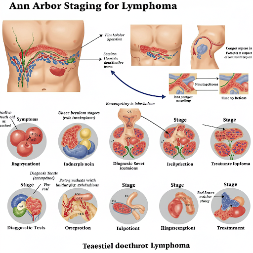
Image: Ann Arbor Staging for Lymphoma
(A diagram mapping out affected lymph nodes in different stages.)
4. Special Staging Considerations
4.1 Pediatric Cancer Staging
- Uses the Children’s Oncology Group (COG) and International Neuroblastoma Staging System (INSS).
- Focuses on tumor spread, risk groups, and response to treatment.
4.2 Colon and Rectal Cancer Staging (Dukes’ Classification)
- Dukes A: Limited to bowel wall.
- Dukes B: Invades through bowel wall but no nodes.
- Dukes C: Lymph node involvement.
- Dukes D: Distant metastasis.
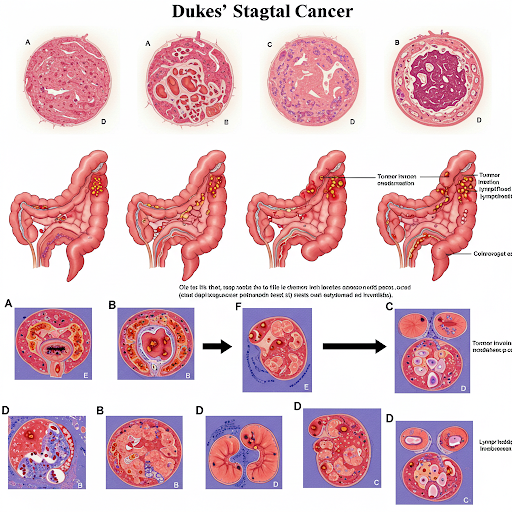
Image: Dukes’ Staging for Colorectal Cancer
(A cross-sectional view of the bowel wall showing cancer progression.)
5. Importance of Cancer Staging
- Guides treatment selection (surgery, chemotherapy, radiotherapy).
- Helps in predicting patient survival (prognosis).
- Standardizes clinical trials and research.
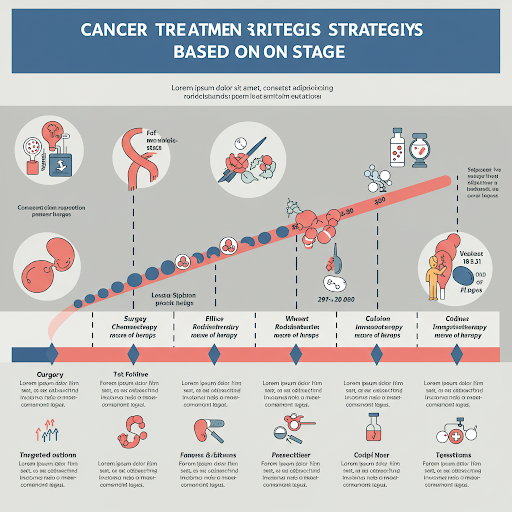
Image: Cancer Treatment Strategies Based on Stage
(A flowchart mapping out treatment options for different stages.)
6. Conclusion
Cancer staging is essential for diagnosis, treatment planning, and prognosis prediction. The TNM system is widely used for solid tumors, while other staging methods like SEER, FIGO, Ann Arbor, and Dukes’ are specific to certain cancers. With advancements in imaging and molecular diagnostics, staging is becoming more precise, improving patient outcomes.